
Shoulder Fracture Overview
A fracture occurs when there is a “break” or a “crack” in the bone. Within the shoulder area, there are 3 distinct bones that could suffer a fracture: the collarbone (clavicle), the upper arm bone (proximal humerus) and the shoulder blade (scapula). A shoulder fracture typically occurs from a sudden force to the arm, such as a hard fall, a blow made during impact through a sport, falling down, or in high-energy situations such as a car accident. Dr. Peter Millett, orthopedic shoulder specialist, treats patients with shoulder fractures from Vail, Aspen and the surrounding Denver, Colorado communities.
What are the symptoms of a shoulder fracture?
A shoulder fracture can be extremely painful to the person suffering from the injury. Some common symptoms associated with a shoulder fracture, a fractured collarbone or a break made to the arm, include:
- Intense shoulder pain
- Swelling of the shoulder area
- Tenderness in the shoulder area
- A bump or disfigurement under the skin at the site of the break
- Bruising or discoloration around the break
- Cracking or mechanical symptoms from the bone edges rubbing on one another
- Inability to move the shoulder or arm without pain
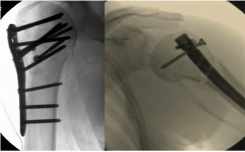
(Click to Enlarge) Left postoperative x-ray image shows a proximal humerus fracture fixed with a plate and screws. Right postoperative fluoroscopic image shows a distal humeral neck fracture fixed with an intramedullary nail.
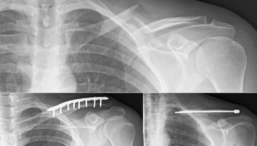
(Click to Enlarge) The top image shows a mid-shaft displaced clavicle fracture. These fractures are typically repaired using either a plate on the superior aspect of the clavicle or an intramedullary pin. The bottom left x-ray shows the superior clavicle plate. The bottom right x-ray shows the intramedullary pin.
How are fractures in the shoulder treated?
There are specific treatment recommendations for each type of shoulder fracture. Clavicle and proximal humerus fractures are frequently treated surgically in active patients when there is significant displacement (separation) of the bone ends. Non-surgical and arthroscopic surgical options are always discussed to make sure the injured bone heals properly. Those are outlined below:
Collarbone Fractures
The clavicle—or collarbone—is the bone on top of one’s chest on both sides at the front of both shoulders. The clavicle is very easy to feel as it represents itself as a prominent bony connection between the shoulder and the body itself. Because the clavicle is located directly under the skin in an area with little soft tissue coverage, fractures to the area not only produce intense shoulder pain but they are also cosmetically obvious to the naked eye. Fractures within the collarbone are among the most common fractures of the shoulder area. Treatment of a collarbone fracture typically does involve surgery when there is significant separation of the bone ends or shortening (overlap) of the bone ends. Surgical treatment is performed with a small pin or plate, depending on the configuration of the fracture fragments, that hold the bone in place until it heals solidly (usually 6 weeks or so). Surgery not only insures that the bone heals in proper alignment but it also results in a better long term functional outcome for the arm, decreases pain in the immediate area around the fracture, allows earlier resumption of everyday activities, allows earlier return to sport in some cases, and decreases the risk of a malunion (healed in the wrong position) which frequently heal with a cosmetically displeasing bump. In some instances, there can be late complications that may need surgical treatment such as when the bone has healed incorrectly or when it has failed to heal. Dr. Millett has done extensive research on this topic and has successful outcomes for his patients who have experienced a clavicle fracture.
When a clavicle fracture is minimally displaced or non-displaced, non-surgical treatment is recommended with plenty of rest. Patients will usually need to wear a sling to help prevent movement and to keep the area immobile. For the most part, these types of collarbone fractures will repair and heal themselves in about 12 weeks.
Upper Arm Bone Fractures
When the region around the upper arm is fractured, the break can take place in 3 areas: near the shoulder joint (called a proximal humerus fracture), within the mid-shaft of the arm (between the shoulder and the elbow commonly referred to as a mid-shaft humerus fracture) and last, near the elbow joint (called a distal humerus fracture). The majority of all fractures within these regions can be corrected with a brace or sling.
Similar to the collarbone, non-surgical methods do not work, or if the injury is too severe, fracture fixation surgery will need to be performed to correct the break or fracture. For instance, if there is a displacement or an overlap of the break greater than 1 centimeter, if the bone points to the skin or protrudes through it, or for patients who desire a quicker return to their normal activities; surgery may then be recommended and necessary to fix the fracture. Fixation can be done with pins, plates and screws, or even prosthetic replacement (called hemiarthroplasty, or reverse total shoulder replacement), depending on the severity of the injury. The fracture fixation procedure may vary depending on the area that is being treated and on the severity of the break.
CLICK IMAGE TO ENLARGE
Minimally-Invasive Shoulder Fracture Repair Surgery
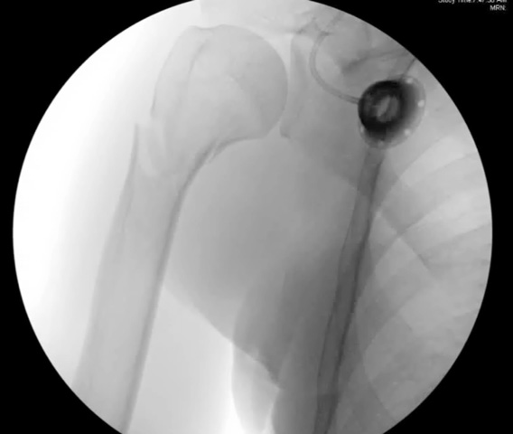
Fractures to the proximal humerus can also be treated using minimally-invasive shoulder fracture repair surgery (arthroscopic and percutaneous). This is pursued when the break or fracture is of a lesser extent and can be done with limited hardware. This type of surgery is generally less painful, less likely to cause complications, and may enable a more rapid recovery process than traditional surgery.
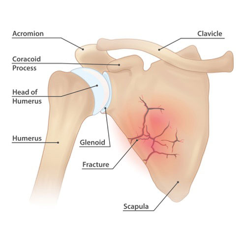
Scapula (Shoulder Blade) Fractures
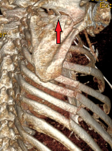
The shoulder blade (known as the scapula) represents the large, flat bone in the upper back area. Fractures of the shoulder blade area are very rare and if a break has occurred in this region, it is usually accompanied by other injuries. The most common cause for this type of an injury is from high-energy trauma—such as a car accident, motorcycle crashes, or extreme falls. This particular type of fracture will need to be evaluated and then a treatment plan set forth. In most cases, a sling immobilization brace can heal the fracture. In cases where significant angulation of the broken bones has occurred, fracture fixation surgery will need to be performed. In some instances there can be late complications form these types of fractures that then require surgery.
CLICK IMAGE TO ENLARGE
CLICK IMAGE TO ENLARGE
Acute Fracture Management
For the fracture conditions listed above in the collarbone, shoulder and arm regions, the following guidelines should be followed while the injury is healing without surgical intervention:
- Apply ice to the injury for 15 to 20 minutes each hour for the first 1 to 2 days. Protect your skin with a light cloth to avoid burning the skin.
- Immobilize the area with a sling.
- Wear your sling until Dr. Millett specifies that you can remove it. You may take off the sling to dress or bathe, but be careful not to move your arm.
- For pain management, Dr. Millett may prescribe pain medication. Please take as prescribed and follow the precautions listed for the drug. You also may take over-the-counter medicines for pain.
You should call our office immediately if you experience the following:
- Intense pain in the collarbone area, shoulder or arm
- Pain that does not gradually diminish
- Excessive swelling in the area of the fracture
- Numbness or cold in the arm or shoulder
- Fever
- Trouble breathing
For more information on shoulder fractures, please contact the office of Dr. Peter Millett.