

CuffMend™ Rotator Cuff Augmentation System
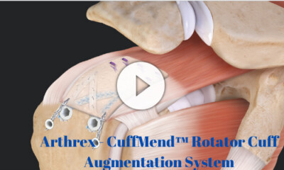
CuffMend™ Rotator Cuff Augmentation System
In this video presentation, Dr. Peter Millett demonstrates how to use the CuffMend™ rotator cuff augmentation system with a full-thickness SpeedBridge™ repair. Dr. Millett highlights fixating the ArthroFlex graft with TissueTak™ tendon anchors and completing the repair augmentation with 3.5 mm PushLock® anchors.
ArthroFLEX® is a registered trademark of LifeNet Health.
Proximal Humerus Fracture: Open Reduction Internal Fixation With SuturePlate™ Humeral Fracture Plating Technique | Orthopedia
Proximal Humerus Fracture: Open Reduction Internal Fixation With SuturePlate™ Humeral Fracture Plating Technique
In this video presentation, Dr. Millett demonstrates an open reduction, internal fixation with SuturePlate™ Humeral Fracture Plating Technique for a proximal humerus fracture.
Distal Clavicle Fracture Repair – Utilizing a Knotless Coracoid Fixation Device
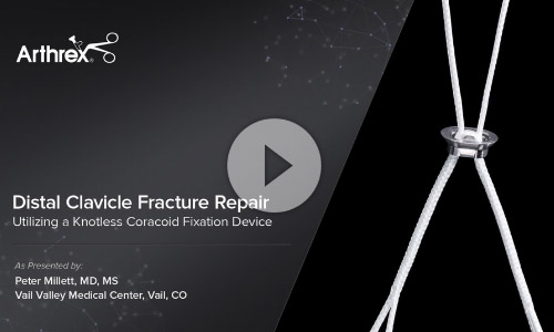
Distal Clavicle Fracture Repair – Utilizing a Knotless Coracoid Fixation Device
Peter Millett, MD, MS, demonstrates arthroscopically assisted coracoid fixation to treat lateral clavicle fractures utilizing a clavicle plate and a knotless clavicle plate button. The plate button is preassembled with a knotless TightRope®‐like suture and is designed to be used with a Dog Bone™ Button and an Arthrex® distal clavicle plate. Dr. Millett shows how the combination of a plate and knotless coracoid fixation can give the surgeon a strong, knotless, low profile repair option to treat difficult lateral clavicle fractures.
Arthroscopic Subacute AC Joint Reconstruction Using the Dog Bone™ Button and Allograft
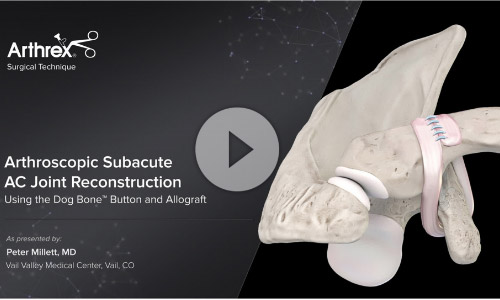
Arthroscopic Subacute AC Joint Reconstruction Using the Dog Bone™ Button and Allograft
Peter Millett, MD, demonstrates his arthroscopic technique for a subacute AC joint reconstruction using the Dog Bone™ Button, FiberTape® and an allograft wrap around the coracoid. He utilizes a new adjustable AC guide to drill 3 mm tunnels in the clavicle and coracoid and uses a Switching Stick and Obturator to facilitate arthroscopic allograft passage around the coracoid.
Expanded SpeedBridge™ for Massive Revision Rotator Cuff Tear Augmented with ArthroFlex® Acellular Dermal Extracellular Matrix
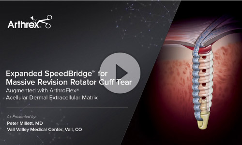
Expanded SpeedBridge™ for Massive Revision Rotator Cuff Tear Augmented with ArthroFlex® Acellular Dermal Extracellular Matrix
Peter Millett, MD, performs a live surgery on a patient with two prior failed cuff repairs. After an arthroscopic evaluation and multiple releases, he determines that the patient's poor tendon quality requires augmentation and decides to perform an open, six-anchor SpeedBridge™ that incorporates a 2 mm thick ArthroFlex®* allograft. He highlights several pearls regarding graft sizing, suture passing and anchor placement to achieve an excellent result.
Arthrex PowerPick™ Microfracture of a Femoral Condyle Lesion
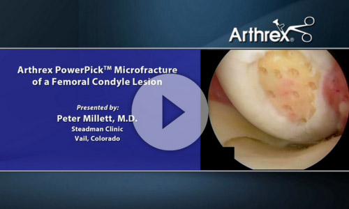
Arthrex PowerPick™ Microfracture of a Femoral Condyle Lesion
In this surgical technique video, Peter Millett, MD, (Vail, CO) discusses the use of the PowerPick in the treatment of an articular cartilage lesion of the femoral condyle.
The PowerPick produces 1.5 mm holes rapidly and in close proximity to one another, helping to prevent damage to surrounding bone and healthy cartilage, making it the ideal tool for treating articular cartilage defects.
Arthrex – Tensionable Knotless Bankart Repair Using the Knotless 1.8 mm FiberTak® Implant System
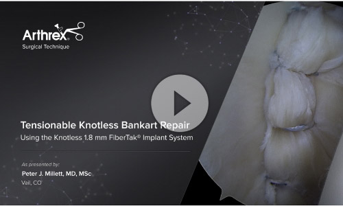
Tensionable Knotless Bankart Repair Using the Knotless 1.8 mm FiberTak® Implant System
Peter J. Millett, MD, MSc, (Vail, CO) performs a cadaveric demonstration of a knotless Bankart repair using Knotless 1.8 FiberTak® suture anchors. Dr. Millett demonstrates how to use the unique adjustability of this knotless suture anchor to achieve an anatomically tensioned soft-tissue glenohumeral repair.
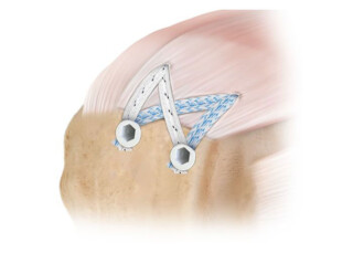
Strength and Reliability of the SpeedBridge™ Repair – Case Presentation
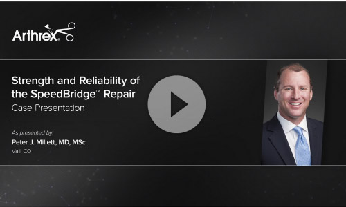
Strength and Reliability of the SpeedBridge™ Repair – Case Presentation
Peter J. Millett, MD, MSc, (Vail, CO) describes his 5‐year results using knotless SwiveLock® suture anchors combined with FiberTape® suture for double‐row SpeedBridge™ rotator cuff repairs. Dr. Millett also shares 2 clinical cases highlighting the resilience of the SpeedBridge repair after his patients experienced significant postoperative shoulder trauma.
SLAP Repair with Knotless SutureTak® Anchors
SLAP Repair with Knotless SutureTak® Anchors
Peter J. Millett, MD, MSc, demonstrates the surgical technique for a SLAP repair using the knotless SutureTak® anchor.
Dr. Millett describes where to place the ports both anteriorly and posteriorly. It is important during this step to not capture the biceps tendon.
He then explains the process for placing the SutureTak® Anchors and which anchor to pull through the port.
He will use a guide to thread the fiber-wire through the labrum and pull through and out the port. Dr. Millett then demonstrates how to keep the anchor threads from tangling. He shows how to make sure an adequate amount of suture is obtained and secured.
Before final tensioning, he will use the tension cutter device to both tension the threads and then cut them appropriately.
The process is repeated with the anterior anchor.
Final amounts of tension are applied, and the cutter is used when completed.
The shoulder labrum is now secured anteriorly and posteriorly without over-tensioning the biceps.
The knotless repair is particularly important to avoid friction in the overhead throwing athlete.
SuturePlate™ Proximal Humeral Fracture Repair
SuturePlate™ Proximal Humeral Fracture Repair
Subpectoral Biceps Tenodesis
Subpectoral Biceps Tenodesis
In this video presentation, Dr. Millett demonstrates a subpectoral biceps tenodesis.
Dr. Millett shows how to mark the incision and where to cut to find the facia and cut through, then locate the short head of the biceps. The assistant will retract the pectoralis major while Dr. Millett locates and retracts the tendon. 2cm up from the muscle/tendon junction, use a whipstitch. Next, open the bicipial groove and take off the soft tissues. Visualize the bone, then use an 8mm tenodesis driver with sofit tissue sleeve. Drill a hole, perpendicular to the bone. Secure the suture thread tendon through the created hole. Advance the screw, then feel that the scres is secure. Finally, tie the sutures over the top so that the tendon is fixed in the bicepital groove.
Return to Sports in the COVID Era: A Community Approach for Youth Athletes
In this video, Dr. Stephanie Pearce discusses the steps that should be taken for young athletes to return to sport in the time of COVID.
Dr. Pearce discusses the following:
- COVID-19 is an everchanging situation with may different possible health anomalies to be aware of.
- A safe and healthy return to sport is important.
- COVID and Sports
- CDC's Risk Assessment in Sports
- What to do if an athlete gets COVID
- COVID and the athlete's heart
- Multisystem Inflammatory Syndrome in Children (MIS-C)
- How is it diagnosed?
- How is it Treated?
- NCAA on Return to Sport
The steps for returning to sport are as follows:
- Step 1: Daily Symptoms Form
- Step 2: Exercise Guidance
- Step 3: Medical Clearance
- Step 4: COVID 19 – Graduated Return to Play
Arthroscopic Pearls using Knotless FiberTak® Suture Anchor for Labral Repair
In this video presentation Dr. Millett demonstrates arthroscopic pearls using the knotless FiberTak® suture anchor for labral repair. The are two cannulas used in this repair, a 5 mm cannula superiorly just below the biceps tendon and an 8.25 mm canula just above the subscapularis. For placement of the first anchor, a 15 degree curved guide was used to get down low on the glenoid face and a flexible drill was utilized to create a drill hole. A 1.8mm knotless FiberTak anchor was started by hand and then tapped into place in the drill hole. A 25 degree tight left lasso was placed around the labrum and a shuttling wire allowed the repair suture to be shuttled out the 8.25 mm cannula. The repair suture is then placed through the looped end of the shuttling suture and folded onto itself before being pulled through the anchor. An arthroscopic grasper is then used to pull up on the tissue to facilitate shift of the tissue from inferior to superior prior to tensioning and securing the tissue in place. The repair suture is then cut right at the cartilage to decrease the risk of tangling with the placement of additional anchors. This process was repeated from inferior to superior until all anchors were placed and sufficient repair was achieved. Labral repair was performed in this case with four suture anchors placed in the anterior glenoid at approximately the 7 o'clock, 8 o'clock, 9:30, and 10:30 positions for this left shoulder demonstration.
Knotless, Tensionable Labral Repair
 Peter J. Millett, MD, MSc, (Vail, CO) describes his experience using the Knotless 1.8 FiberTak® suture anchor for instability procedures. Dr. Millett also shares biomechanical testing and clinical results with the Knotless 1.8 FiberTak suture anchor.
Click here to view: Knotless, Tensionable Labral Repair
Tensionable Knotless Bankart Repair Using the Knotless 1.8 mm FiberTak® Implant System
 Peter J. Millett, MD, MSc, (Vail, CO) performs a cadaveric demonstration of a knotless Bankart repair with the Knotless 1.8 mm FiberTak® suture anchors. Dr. Millett demonstrates how to use the unique adjustability of this knotless suture anchor to achieve an anatomically tensioned soft tissue glenohumeral repair.
Click here to view: Tensionable Knotless Bankart Repair Using the Knotless 1.8 mm FiberTak® Implant System
Posterior Labral Repair Using Short 2.9 mm PushLock® and LabralTape™
 Peter Millett, MD, (Vail, CO) performs a live surgery using knotless 2.9 mm BioComposite™ PushLock® suture anchors with LabralTape™ to complete a low profile posterior labral repair. He discusses the benefits of knotless fixation in minimizing the postoperative trauma that can be caused by prominent knot stacks. He also highlights the use of LabralTape for increased tissue holding capability over standard sutures.
Click here to view: Posterior Labral Repair Using Short 2.9 mm PushLock® and LabralTape™
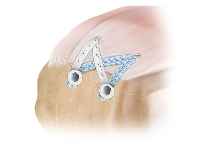
SpeedBridge™ Rotator Cuff Repair Using the SpeedBridge™ Implant System
 Peter Millett, MD, (Vail, CO) performs a live surgery using knotless BioComposite™ SwiveLock® suture anchors with FiberTape® to complete a quick and secure double-row rotator cuff repair that restores the rotator cuff footprint and maximizes contact between the tendon and bone. Dr. Millett prefers a totally knotless construct to help evenly spread the load across the entire construct. He also highlights use of the SutureLasso™ for retrograde suture passage and uses a PowerPick™ to prepare a bleeding bone bed in the rotator cuff footprint.
Click here to view: SpeedBridge™ Rotator Cuff Repair Using the SpeedBridge™ Implant System
Rotator Cuff Disorders and their Treatment
 Dr. Peter Millett is an orthopaedic surgeon and partner at the Steadman Clinic, and is consistently selected as one of the "Best Doctors in America." In this course, he covers anatomy, evaluation, and rehabilitation of common rotator cuff disorders. He will take you through the characteristics of various types of rotator cuff tears and other pathologies, outline keys to success and identify the different prognostic factors that contribute to good outcomes with rotator cuff patients. You will learn how to evaluate and examine a patient with a possible rotator cuff pathology, and the different repair techniques will be examined from an evidence-based perspective. Dr. Millett concludes with a presentation of the three-phase approach to the rehabilitation of rotator cuff patients.
Register Now: Rotator Cuff Disorders and their Treatment